1. You will be instructed to taper your oral steroid agent over a course of several days. As long as you are on Decadron, you will continue to take Pepcid or Zantac to prevent gastritis.
  1. You will continue with the prescribed anti-epileptic medication until instructed to discontinue.
  1. Your postoperative headache will improve very fast, and this will be at its minimal, or very manageable by with an oral pain medication, by the time you go home.
  1. You will be asked to return 10-14 days following your surgery for suture removal.
  1. You will use your common sense as to how much or little you do at home. Doing too much will wear you out and at times cause worsening headaches, while doing too little or being sedentary at home may cause dangerous complications such as pneumonia and deep venous thrombosis (DVT) in your calves.
  1. My recommendation is to be as active as you can tolerate, going for walks and increasing the overall level of activity on a daily basis.
  1. Expect to return to work in 4-6 weeks following surgery in most situations.
  1. You will again return for a routine postoperative follow-up evaluation in 6-8 weeks to see me.
  1. For patients with a benign meningioma (WHO Grade I), your follow-up MRI will be 1 year after surgery.
  1. For patients with an aggressive meningioma (WHO Grade II), your follow-up MRI will be 4-6 months after surgery.
  1. For patients with a malignant meningioma, you will be arranged to have radiation treatment(s) shortly following surgery. The follow-up MRI will be in 3 months after completion of radiation treatments.


  1. First of all, in preparation for surgery, you will be instructed to discontinue any blood thinners (such as Aspirin, Advil Coumadin, Plavix, etc.) from 3 to 7 days prior to surgery.  If this instruction is not followed, your surgery will be canceled.
  2. You will be instructed to not eat or drink anything after midnight prior to the morning of surgery.  On the day of surgery, you may take important medications with minimal sips of water (such as medications for hypertension).
  3. You will be asked to come into the hospital 90 minutes to 2 hours prior to your scheduled surgery.
  4. Just prior to being taken into the operating room (OR), you will meet your surgeon again to go over any last minute questions or concerns you may have, and also meet your anesthesiologist.
  5. Once in the OR, your anesthesiologist and the OR nursing personnel will do a “time out” to confirm your identification and the scheduled surgery to be performed, followed by  preparing you for general anesthesia and surgery, including starting 2 IV lines, an arterial line if needed and a Foley catheter if needed (for bladder emptying during surgery).  Then, you will “go to sleep” after having an endotracheal tube inserted.
  6. Surgery will then begin by the surgeon with positioning of the body and the head appropriate for the particular surgery you are having.  For most brain operations, your head will be fixed in a 3-pin frame for the duration of your surgery.
  7. After optimal body/head positioning, an area on the scalp around the planned incision will be shaved (as little as possible), and the surgical site prepped with sterilizing solution.
  8. Then, the head and body will be “draped” with only the shaved & prepped planned incision area of the scalp exposed.
  9. Surgery then begins after another “time out”, to confirm that the correct patient is in the OR, and the correct surgery is being performed on the appropriate side of the head/brain as scheduled.
  10. Surgery will last anywhere from about 1 hour (for small, convexity meningiomas) to 4-6 hours (for large, complex skull base meningiomas).
  11. After surgery, a sterile head dressing is applied, general anesthesia reversed, and the endotracheal tube removed as you “wake up” from surgery and anesthesia.
  12. A quick neurological evaluation is usually performed by the surgeon after you “wake up” from surgery, prior to leaving the OR.
  13. You will be taken either to the recovery room (“PACU”), followed by a nursing floor or directly to the intensive care unit (“ICU”) for postoperative management.
  14. Your will meet your family about an hour or two after completion of your surgery in your room.
  15. You will obtain a postoperative MRI on the day following your surgery to evaluate the extent of surgery.
  16. You will be encouraged to get out of bed and move around from the day following surgery when your diet will also be advanced, as tolerated, to a regular diet.
  17. Your hospital stay will vary anywhere from 1 to 3 days (for most meningioma surgery).


  1. For those with significant symptoms caused by “peritumoral edema”, i.e. swelling around the meningioma, it is better to reduce/minimize this swelling prior to surgery. You will be treated with approximately a 1-3 week course of an oral steroid agent called, “Dexamethasone (or Decadron)”.  The length of steroid course is determined by the severity of edema and symptoms.
  1. Oral steroids may cause stomach irritation (“gastritis”), and to prevent this from occurring, you will be started on either Pepcid or Zantac along with your Decadron.
  1. Depending on the tumor size, location and symptoms, you will also be started on a seizure medicine (“anti-epileptic”) for seizure prevention, usually Keppra or Dilantin, which will be discontinued about 4-6 weeks following surgery provided that you remain seizure-free after surgery.
  1. If your symptoms are not debilitating, you will work or carry on with your normal daily activities until the day of surgery.
  1. In the few weeks leading up to your surgery, you will obtain the following preoperative evaluations: Chest X-ray, EKG, blood tests (Metabolic panel, CBC, PT/INR, PTT)
  1. In preparation for surgery, you will be instructed to discontinue any blood thinners (such as Aspirin, Advil, Coumadin, Plavix, etc.) from 3 to 7 days prior to surgery. If this instruction is not followed, your surgery will be canceled.
  1. You will be instructed to not eat or drink anything after midnight prior to the morning of surgery. On the day of surgery, you may take important medications with minimal sips of water (such as medications for hypertension).


  1. Meningiomas are mostly (92%) benign tumors arising from the brain covering (“the meninges”).
  1. Meningiomas are the most common “primary” brain tumors with the annual incidence approaching 80/million (when including “incidental” tumors).
  1. Meningiomas are more common in the female sex by 2-2.5:1.
  1. Meningiomas most commonly affect the middle aged population, with the average age at the time of initial diagnosis being 54 years.
  1. Meningiomas are most likely caused by alterations or damages that have occurred in the patient’s genes. Unfortunately, however, exactly which gene(s) lead to meningioma formation, other than the NF2 gene in select patients, and why those genetic changes occurred are unknown as of today.
  1. Meningiomas grow slowly, with 44% of observed tumors showing growth within 4 years.
  1. Not all meningiomas require treatment at the time of initial diagnosis.
  1. Management options include observation, surgery and radiation/radiosurgery.
  1. Surgery is the treatment of choice for most patients with meningiomas.
  1. Surgery is recommended for young, healthy, symptomatic patients and those with large tumors.
  1. The factors determining long term survival include the tumor histology (graded according to the WHO classification of Grades I (“benign”) vs. II (“aggressive”) vs. III (“malignant”)) and the extent of resection, which commonly is, in turn, influenced by the surgeon’s experience.
  1. Gross tumor removal along with removal of the surrounding/involved dura and bone may lead to surgical “cure” in benign meningiomas.
  1. The factors determining patient’s outcome following surgery include the patient’s co-morbidity (C), tumor location (L), patient’s age (A), tumor size (S), symptoms (S), in addition to history of any prior surgery or radiation, as well as the surgeon’s experience.
  1. Young, healthy patients with small and/or asymptomatic meningiomas undergoing surgery by an experienced surgeon do very well following their surgery.


Meningioma Learning Curve Outcome

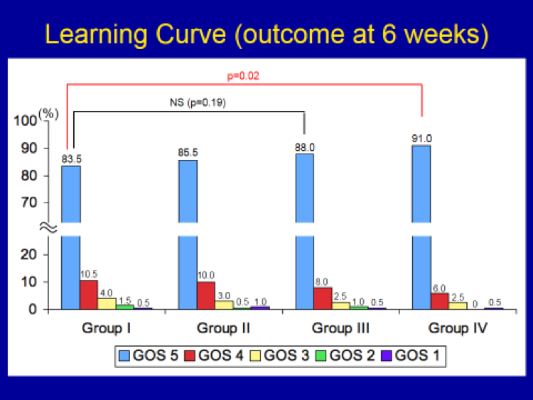
In order to see how the surgeon’s (i.e. my) experience affected outcome following meningioma surgery over time, 800 meningiomas were subdivided into groups of 200 each.  The following time frame was required to accumulate the experience of 200 cases each:

Group I:  7 years

Group II:  3 years and 3 months

Group III: 2 years and 6 months

Group IV: 2 years and 6 months

There was a gradual increase in the incidence of “Normal” outcome group in each of the successive groups of 200 cases, going from 83.5 % in Group I, eventually to 91 % in Group IV.  However, this difference (i.e. improvement) was not statistically significant until the last group (Group IV) of 200 cases.  In other words, it takes a surgeon at least 600 cases of accumulated experience to make a statistically significant improvement in outcome in meningioma surgery.  In short, surgeon’s experience is a critically important factor in determining outcome in meningioma surgery.               


Meningioma Previous Radiation-outcome

Previous radiation:  In order to study whether a history of radiation influences outcome in meningioma surgery, “Radiation (Yes)” group was compared to those with no prior radiation with respect to outcome (GOS) (based on a review of 800 operative cases, personal series).  A total of 93 patients had previous radiation.  Many of these patients also had prior operations.  

The incidence of “Good” outcome (GOS 4 & 5) patients goes down significantly from the “No radiation” group to the “Prior (Yes) radiation” group.

Prior radiation, similar to previous surgery, causes scar tissue formation around the tumor, making exposure and identification of the tumor and the surrounding “normal” anatomic structures difficult and, at times, dangerous, adding to overall surgical risks.  Patients with a history of previous radiation do less well following surgery.


Meningioma Previous Surgery Outcome

Previous surgery:  To see whether or not a history of prior surgery had any influence on outcome following meningioma surgery, “Previous surgery (Yes)” group was compared to those with no prior surgery with respect to outcome (GOS) (based on a review of 800 operative cases, personal series).  A total of 93 patients had previous operation(s).

The incidence of “Good” outcome (GOS 4 & 5) patients goes down significantly from the “No surgery” group to the “Prior (Yes) surgery” group.

In brain surgery, prior operation causes scar tissue formation at the site of surgery, making exposure and identification of the tumor and the surrounding “normal” anatomic structures difficult and, at times, dangerous, adding to overall surgical risks.  Patients with a history of previous surgery do less well following surgery.


Meningioma Symptoms Outcome

Symptoms:  Symptomatic tumors were compared against those causing no symptoms (“Asymptomatic”) with respect to outcome following surgery (based on a review of 800 operative cases, personal series):

Asymptomatic:  Tumors causing no symptoms (i.e. “incidental” tumors)

Symptomatic:  Tumors producing significant symptoms or neurological deficits

The incidence of “Good” outcome (GOS 4 & 5) patients goes down significantly from the “Asymptomatic” tumor group to the “Symptomatic” group.  In particular, the incidence of “normal” outcome is about 98% in the “Asymptomatic” group as compared to 81% in the “Symptomatic” group.  In short, whether or not the tumor is causing any preoperative symptoms does influence how well patients do following their surgery.  This may have to do the the larger size and the presence of peritumoral edema (swelling) in the “Symptomatic” group of patients.  In short, patients with “asymptomatic” meningiomas do extremely well following surgery.


Meningioma Size Outcome

Size of the tumor was assessed with respect to outcome (GOS) by dividing into 3 groups (based on a review of 800 operative cases, personal series):

Small:  Less than or equal to 2 cm in largest diameter

Medium:  Between 2.1 and 4 cm in largest diameter

Large:  Larger than 4 cm in largest diameter   

The incidence of “Good” outcome (GOS 4 & 5) patients goes down significantly, again in a step-wise manner, from the “Small” tumor group to the “Medium” group, then from the “Medium” group to the “Large” group.   No “Poor” outcome was seen in the “Small” group.  In short, the tumor size also does matter in how well patients do following meningioma surgery.  Patients with “small” meningiomas do extremely well following surgery.


Meningioma Age Outcome

Age was assessed with respect to outcome (GOS) by dividing into 3 groups (based on a review of 800 operative cases, personal series):

Group I:  Age less than 60

Group II:  Age between 61 and 70

Group III:  Age greater than 70 

The incidence of “normal” patients (GOS 5) goes down significantly from the Group I to II, then from II to III.   Young patient (<60) do very well from meningioma surgery, while older patients fared less optimally.  In short, age also does matter in how well patients fare following meningioma surgery.


© 2024 Valley Neurosurgical Institute