1. Correct diagnosis, based on a careful analysis of the clinical history and examination, is critical to therapeutic success.
  2.  MRI must be done to rule out the presence of:

MS

Mass lesion (tumor or cyst)

Vascular compression

  1. All trigeminal neuralgia patients should undergo a trial of medical therapy.
  1. Surgery is to be considered when medical therapy fails, or the patient experiences significant side-effects.
  1. Approximately 50% of patients will eventually require surgery due to failed medical therapy.
  1. TN patients should be aware of all – ablative vs. non-ablative – surgical options.
  1.   For non-MS patients with evidence of vascular compression on the MRI scan, MVD is  the treatment of choice:

Surgery is simplified (minimally invasive surgery)

4-cm incision,

<2-cm bone opening (“suboccipital craniectomy”)

45-minute surgery

Foley catheter not needed (for bladder drainage during surgery)

Minimal blood loss, avoiding blood transfusion

1-2 days of hospital stay

Negligible permanent facial numbness as compared  to 80% following ablative surgery


MVD Positioning

The patient is positioned lying flat (“supine position”) with a small area behind the right ear shaved, and the incision marked.


MVD Skin Incision

A 4 to 4.5 cm-incision is marked (1 ¾”) behind the right ear.


MVD Suboccipital Craniectomy

The size of bone opening (“craniotomy” or “craniectomy”) shown above is 15 mm, which is less than the diameter of a dime (18 mm).


MVD Dural Opening and Approach to the Trigeminal Nerve

Exposure of the right trigeminal nerve (“N”) and the surrounding vein (“V”) and artery (unmarked, on the far side between the vein and nerve).


MVD Exposure of the Trigeminal Nerve

Surgical instruments – suction on the left and bipolar electro-cautery forceps on the right – being used through the bone opening.


MVD Exposure of the Trigeminal Nerve

The offending artery (“A”) is being dissected away from the nerve (“N”) with a micro-instrument (appearing blurry, just below the letter “A” marking the offending artery).


MVD Separation of Artery from Trigeminal Nerve

The offending artery is completely separated far away from the nerve.


MVD Placement of a Teflon Felt

A Teflon felt is placed between the trigeminal nerve and the surrounding artery (now covered by the felt), completely relieving pressure off of the trigeminal nerve, thereby completing the surgery of “microvascular decompression” (“MVD”).


Surgery is considered when (1) medical therapy fails to adequately control trigeminal neuralgia, (2) patients are unable to tolerate medications’ side-effects, or (3) patients do not like the prospect of being on long-term medications. 

Surgical Options:

A) Ablative (“to destroy a function by application of a noxious substance”) Surgery:

For some unknown reason, when a segment of the trigeminal nerve is “damaged” (by way of “lesioning” or “ablation” applied to it) by a noxious agent, trigeminal neuralgia pain disappears or improves.  There are several surgical means of creating a focal area of “ablation” to the trigeminal nerve, and these include:

Gamma Knife (GK) Radiosurgery

A single shot, focused radiation targeted to the trigeminal nerve

Balloon Compression Rhizotomy

Lesioning by way of a mechanical compression to the nerve

Glycerol Rhyzotomy

Chemical lesioning to the nerve by a type of alcohol

Radiofrequency (RF) Rhizotomy

Thermal (heat) lesioning to the nerve by RF

Long-term success rates:  Glycerol 60%, Gamma Knife 65%, Balloon 67%, RF 75%

Advantages:

Short operative time (15 to 45 minutes)

Avoids general anesthesia, performed under IV sedation/local anesthetic

Good initial pain relief

Same-day procedure and quick recovery

Disadvantages:

Permanent facial numbness – RF 80%, Balloon 50%, Glycerol & GK 30%

Significant pain during surgery (performed without general anesthesia)

Need to repeat procedures due to relatively high pain recurrence rates

      Indications for Ablative Surgery:  (When to proceed with ablative surgery?) 

  1. Trigeminal neuralgia patients with MS (multiple sclerosis).
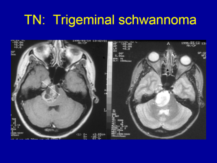
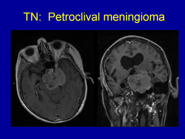
  2. Trigeminal neuralgia patients with no clear vascular compression on the preoperative MRI.
  3. Trigeminal neuralgia patients with significant medical illnesses which preclude major surgery under general anesthesia.
  4. Patient’s personal choice after thorough discussion of all options.
  5. Which specific ablative procedure to be done depends on the individual surgeon’s personal experience and choice.


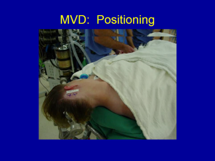
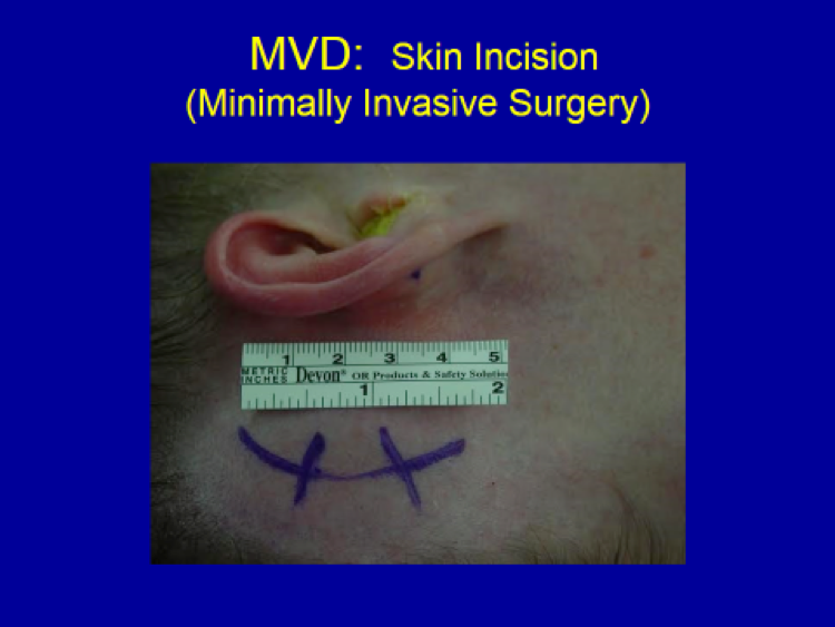
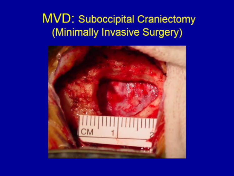
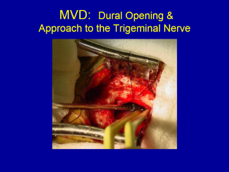
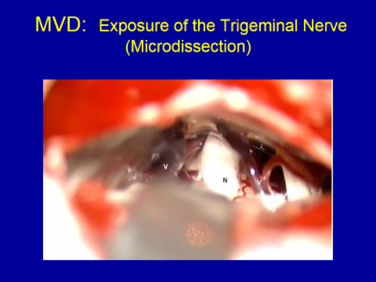
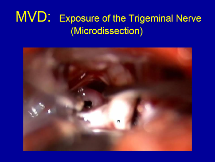
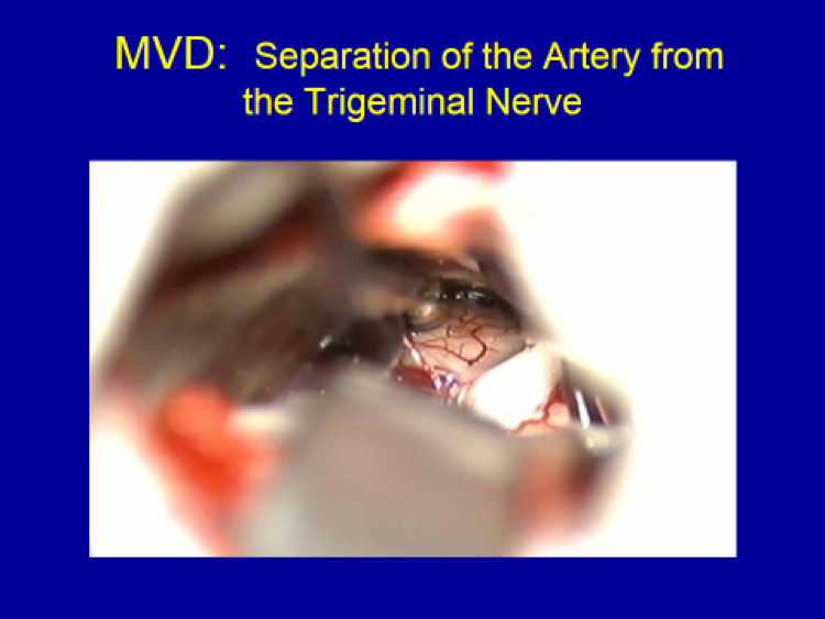
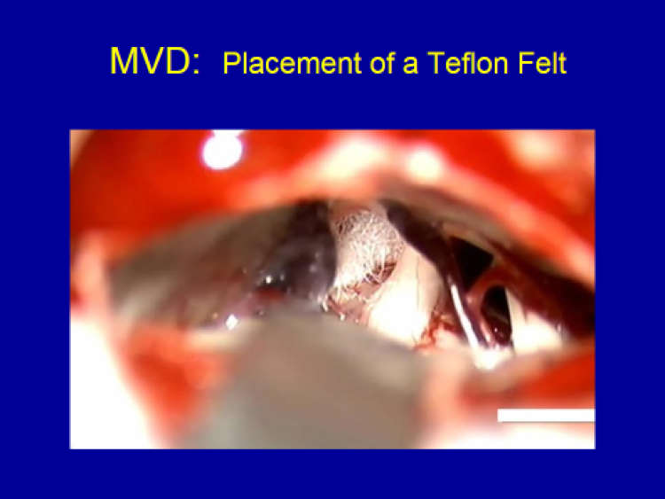
B) Non-Ablative Surgery: Microvascular Decompression (MVD)        

The only non-ablative surgical technique available for trigeminal neuralgia patients is microvascular decompression (MVD).

Indications for MVD: 

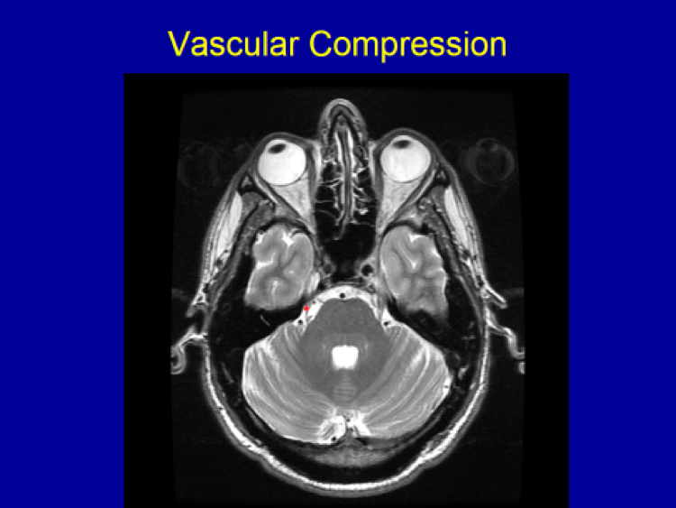
  1. Trigeminal neuralgia patients with clear vascular compression on preoperative MRI.
  2. Patients healthy enough to undergo 1 hour of surgery under general anesthesia.
  3. Patient’s personal choice after discussion of all treatment options.

Advantages:

High success rate (95% when patients are carefully selected)

Low risk of permanent facial numbness

Minimal complications (when surgeon is experienced)

45-minutes of operative time, avoiding Foley catheter or blood transfusion

Small incision and craniotomy (bone opening)

Fast recovery (1-2 days of hospital stay) 

Disadvantages:

            “Brain surgery” with its associated risks (very small, but present)

            Requires general anesthesia (<1 hour)

Personal Recommendation:  Based on treating trigeminal neuralgia patients for nearly 30 years, my personal recommendation is MVD as it guarantees a high success rate with a low risk of permanent facial numbness.  (95% success rate initially, 75-80%  long-term success rate, <5% permanent numbness)


When evaluating a new patient with trigeminal neuralgia, MRI with and without Gadolinium is done to make certain that there is no underlying mass lesion along the course of the trigeminal nerve (e.g. meningioma, schwannoma, epidermoid, or very rarely, skull base cancer).  At the same time, a special MRI protocol (“CISS” or “FIESTA”) is performed to look for any possible “vascular compression” on the trigeminal nerve.


Trigeminal Schwannoma

35 year old female patient’s MRI showing a large tumor arising from the trigeminal nerve (trigeminal schwannoma) as the cause of her trigeminal neuralgia.


Petroclival Meningioma

72 year old female patient’s MRI showed a massive tumor (meningioma) causing severe compression on the trigeminal nerve causing trigeminal neuralgia, in addition to other symptoms (R sided weakness, walking difficulty and headache).


Vascular Compression

42 year old patient’s MRI shows the trigeminal nerve (*) compressed by the adjacent arterial loop. Due to the compression, the trigeminal nerve appears thinner than the trigeminal nerve on the opposite side. Additionally, the nerve is “bowed” compared to the nerve that is normal and straight on the opposite side.


Definition

  1. Sudden, severe and recurring (i.e. “paroxymsmal”) facial pain in the trigeminal nerve distribution (the cranial nerve that supplies sensation to the face including the eye).
  2. Trigeminal neuralgia pain is most often described by patients as “sharp”, “intense”, “lancinating”, or “electrical shock-like”.
  3. Trigeminal neuralgia pain is often characterized as one of the most painful conditions known to mankind.


Diagnosis

The diagnosis of trigeminal neuralgia is made after assessing the patient’s thorough clinical history.  Typical trigeminal neuralgia pain characteristics include:

  1. Paroxysmal
  2. Intermittent (“on & off”), not constant
  3. “Sharp”, “lancinating”, “electrical shock-like” pain
  4. Each shock lasting a split second to 60-90 seconds

Additional diagnostic considerations:

  1. After a series of repetitive shocks of various duration, often there is a prolonged burning/deep ache lasting minutes to hours.
  2. Pain characteristics may be altered by trigeminal neuralgia medications or prior surgical procedures.
  3. “Triggering factors” are often present, which provoke typical trigeminal neuralgia pain. These include touching the sensitive areas (“triggering zones”), wind blowing on the face, drinking/eating, smiling, brushing teeth, putting on make-up, etc.


Differential Diagnosis (“What are other diagnostic possibilities?”):

Glossopharyngeal neuralgia

Geniculate neuralgia

Postherpetic neuralgia

Sphenopalatine neuralgia

TMJ

Cluster headache

Atypical facial pain

Trigeminal neuropathic pain (post-traumatic or idiopathic)

Facial pain of the dental, orbital or nasal sinus origin

(e.g. Root canal, post-surgical, infection, cancer)


I have always felt a great sense of gratitude towards all my former patients who taught me so much about the profession I love.  Two of the most important personal principles of mine in medicine (& neurosurgery) are (1) to treat each patient as family, and (2) to regard each patient as a teacher.  It is this second principle that motivated me throughout my career to conduct research studies, write academic papers, and learn from each of my patients so that my future patients will receive better and improved care.  This handbook is a small token of my deep appreciation to all my patients – my real teachers.  As you read this Handbook, please take a moment to thank those who came before you, without whom this Handbook and the improved care I am now able to provide today would not be possible.

It is my wish and intention that this handbook will give trigeminal neuralgia patients enough basic information about their newly diagnosed condition so that their anxiety is eased, and they are better equipped to make educated treatment decisions.

It is also my great wish that the facts outlined in this handbook will give them hope.  Rather than asking “Why me?” as they suffer through excruciating pain, I want all those who read this to finish the handbook on a positive note, having hope that they indeed have excellent treatment options available to them that will result in relief of this awful condition.

With best wishes,

Joung H. Lee, M.D.
President, Valley Neurosurgical Institute &
Co-Director, Hollywood Presbyterian Neuroscience Institute
www.ValleyNI.com


  1. You will be instructed to taper your oral steroid agent over a course of several days. As long as you are on Decadron, you will continue to take Pepcid or Zantac to prevent gastritis.
  1. You will continue with the prescribed anti-epileptic medication until instructed to discontinue.
  1. Your postoperative headache will improve very fast, and this will be at its minimal, or very manageable by with an oral pain medication, by the time you go home.
  1. You will be asked to return 10-14 days following your surgery for suture removal.
  1. You will use your common sense as to how much or little you do at home. Doing too much will wear you out and at times cause worsening headaches, while doing too little or being sedentary at home may cause dangerous complications such as pneumonia and deep venous thrombosis (DVT) in your calves.
  1. My recommendation is to be as active as you can tolerate, going for walks and increasing the overall level of activity on a daily basis.
  1. Expect to return to work in 4-6 weeks following surgery in most situations.
  1. You will again return for a routine postoperative follow-up evaluation in 6-8 weeks to see me.
  1. For patients with a benign meningioma (WHO Grade I), your follow-up MRI will be 1 year after surgery.
  1. For patients with an aggressive meningioma (WHO Grade II), your follow-up MRI will be 4-6 months after surgery.
  1. For patients with a malignant meningioma, you will be arranged to have radiation treatment(s) shortly following surgery. The follow-up MRI will be in 3 months after completion of radiation treatments.


  1. First of all, in preparation for surgery, you will be instructed to discontinue any blood thinners (such as Aspirin, Advil Coumadin, Plavix, etc.) from 3 to 7 days prior to surgery.  If this instruction is not followed, your surgery will be canceled.
  2. You will be instructed to not eat or drink anything after midnight prior to the morning of surgery.  On the day of surgery, you may take important medications with minimal sips of water (such as medications for hypertension).
  3. You will be asked to come into the hospital 90 minutes to 2 hours prior to your scheduled surgery.
  4. Just prior to being taken into the operating room (OR), you will meet your surgeon again to go over any last minute questions or concerns you may have, and also meet your anesthesiologist.
  5. Once in the OR, your anesthesiologist and the OR nursing personnel will do a “time out” to confirm your identification and the scheduled surgery to be performed, followed by  preparing you for general anesthesia and surgery, including starting 2 IV lines, an arterial line if needed and a Foley catheter if needed (for bladder emptying during surgery).  Then, you will “go to sleep” after having an endotracheal tube inserted.
  6. Surgery will then begin by the surgeon with positioning of the body and the head appropriate for the particular surgery you are having.  For most brain operations, your head will be fixed in a 3-pin frame for the duration of your surgery.
  7. After optimal body/head positioning, an area on the scalp around the planned incision will be shaved (as little as possible), and the surgical site prepped with sterilizing solution.
  8. Then, the head and body will be “draped” with only the shaved & prepped planned incision area of the scalp exposed.
  9. Surgery then begins after another “time out”, to confirm that the correct patient is in the OR, and the correct surgery is being performed on the appropriate side of the head/brain as scheduled.
  10. Surgery will last anywhere from about 1 hour (for small, convexity meningiomas) to 4-6 hours (for large, complex skull base meningiomas).
  11. After surgery, a sterile head dressing is applied, general anesthesia reversed, and the endotracheal tube removed as you “wake up” from surgery and anesthesia.
  12. A quick neurological evaluation is usually performed by the surgeon after you “wake up” from surgery, prior to leaving the OR.
  13. You will be taken either to the recovery room (“PACU”), followed by a nursing floor or directly to the intensive care unit (“ICU”) for postoperative management.
  14. Your will meet your family about an hour or two after completion of your surgery in your room.
  15. You will obtain a postoperative MRI on the day following your surgery to evaluate the extent of surgery.
  16. You will be encouraged to get out of bed and move around from the day following surgery when your diet will also be advanced, as tolerated, to a regular diet.
  17. Your hospital stay will vary anywhere from 1 to 3 days (for most meningioma surgery).


  1. For those with significant symptoms caused by “peritumoral edema”, i.e. swelling around the meningioma, it is better to reduce/minimize this swelling prior to surgery. You will be treated with approximately a 1-3 week course of an oral steroid agent called, “Dexamethasone (or Decadron)”.  The length of steroid course is determined by the severity of edema and symptoms.
  1. Oral steroids may cause stomach irritation (“gastritis”), and to prevent this from occurring, you will be started on either Pepcid or Zantac along with your Decadron.
  1. Depending on the tumor size, location and symptoms, you will also be started on a seizure medicine (“anti-epileptic”) for seizure prevention, usually Keppra or Dilantin, which will be discontinued about 4-6 weeks following surgery provided that you remain seizure-free after surgery.
  1. If your symptoms are not debilitating, you will work or carry on with your normal daily activities until the day of surgery.
  1. In the few weeks leading up to your surgery, you will obtain the following preoperative evaluations: Chest X-ray, EKG, blood tests (Metabolic panel, CBC, PT/INR, PTT)
  1. In preparation for surgery, you will be instructed to discontinue any blood thinners (such as Aspirin, Advil, Coumadin, Plavix, etc.) from 3 to 7 days prior to surgery. If this instruction is not followed, your surgery will be canceled.
  1. You will be instructed to not eat or drink anything after midnight prior to the morning of surgery. On the day of surgery, you may take important medications with minimal sips of water (such as medications for hypertension).


  1. Meningiomas are mostly (92%) benign tumors arising from the brain covering (“the meninges”).
  1. Meningiomas are the most common “primary” brain tumors with the annual incidence approaching 80/million (when including “incidental” tumors).
  1. Meningiomas are more common in the female sex by 2-2.5:1.
  1. Meningiomas most commonly affect the middle aged population, with the average age at the time of initial diagnosis being 54 years.
  1. Meningiomas are most likely caused by alterations or damages that have occurred in the patient’s genes. Unfortunately, however, exactly which gene(s) lead to meningioma formation, other than the NF2 gene in select patients, and why those genetic changes occurred are unknown as of today.
  1. Meningiomas grow slowly, with 44% of observed tumors showing growth within 4 years.
  1. Not all meningiomas require treatment at the time of initial diagnosis.
  1. Management options include observation, surgery and radiation/radiosurgery.
  1. Surgery is the treatment of choice for most patients with meningiomas.
  1. Surgery is recommended for young, healthy, symptomatic patients and those with large tumors.
  1. The factors determining long term survival include the tumor histology (graded according to the WHO classification of Grades I (“benign”) vs. II (“aggressive”) vs. III (“malignant”)) and the extent of resection, which commonly is, in turn, influenced by the surgeon’s experience.
  1. Gross tumor removal along with removal of the surrounding/involved dura and bone may lead to surgical “cure” in benign meningiomas.
  1. The factors determining patient’s outcome following surgery include the patient’s co-morbidity (C), tumor location (L), patient’s age (A), tumor size (S), symptoms (S), in addition to history of any prior surgery or radiation, as well as the surgeon’s experience.
  1. Young, healthy patients with small and/or asymptomatic meningiomas undergoing surgery by an experienced surgeon do very well following their surgery.

© 2024 Valley Neurosurgical Institute