Observation:  Indications (When to observe?)

  1. Patients with no symptoms (“incidental meningiomas”) of all ages
  1. Patients with mild symptoms in the elderly or those patients with serious medical conditions in whom surgery carries a significant risks.
  1. Patients with a long history of mild or non-progressive symptoms with tumors in the “high risk” location (e.g. hearing loss on one side)
  1. Patients who strongly oppose surgery after thorough counselling

Radiation:  Indications (When to radiate?)

  1. Patients who have residual tumors with documented growth, or with malignant tumor histology (i.e. WHO Grade III)
  1. Patients with nonoperated tumors or recurrence not amenable to safe surgery (due to the patient’s medical condition, tumor location, or age)
  1. Patients who strongly favor radiation over surgery after thorough discussion of all treatment options
  1. Conventional radiation (or IMRT – ‘intensity modulated radiation therapy”) for patients with tumor size greater than 3-4 cm
  1. Radiosurgery (“Gamma Knife Radiosurgery” or “Cyber Knife Radiosurgery”) for patients with tumor size less than 3 cm


  1. As indicated in the preceding “Natural History” Section, not all meningiomas require any treatment, either in the form of surgery or radiation, at the time of initial diagnosis/presentation.  In my personal practice while at the Cleveland Clinic, among over 200 new patients with meningiomas evaluated each year, about 100 patients were treated with surgery, 10 patients with radiation (or radiosurgery) and 100 patients were observed.
  2. Treatment offered (either surgery or radiation) must guarantee an outcome that is far superior over the natural history (i.e. untreated) itself.
  3. Treatment benefits must far outweigh the risks.
  4. There are only two treatment benefits in meningioma management:
    1. Significant alteration of the natural history of the disease (“prolonging life”)
    2. Reversal or improvement of the presenting neurological symptoms/deficits (“improving life quality”)
  5. There are 3 categories of management options:
    1. Observation
    2. Surgery
    3. Radiation/Radiosurgery

Additional reading:

Lee JH & Sade B:  Management Options and Surgical Principles – An Overview (JH Lee, ed), pp. 203-207), Springer, London, 2008


The natural history of a disease (e.g. a meningioma) is the course an illness takes without any intervention (i.e. any treatment).  In other words, the natural history of a meningioma is how that particular tumor behaves in an individual patient when it is just observed without any treatment.

In any given disease, when and what treatment should be administered must be based on thorough knowledge of (1) the disease’s natural history, (2) the risks of the treatment, and (3) the benefits of the treatment.  In surgical management of a meningioma patient, benefits of surgery must far outweigh the risks, and simultaneously, surgery must significantly alter the natural history, either by “curing” the meningioma when “complete” removal is achieved, or by significantly “delaying the (tumor) progression” when “partial” removal is performed, thereby prolonging the patient’s overall survival.

The literature information on meningioma’s “natural history” has been scant, and when available, the past studies were limited to the sample size of less than 100 patients.  My group at the Cleveland Clinic published the largest “natural history” paper on meningiomas in 2011.  In that study, we observed 273 meningiomas in 244 patients for a mean period of nearly 4 years.   In that time frame, 44% of the meningiomas showed growth.  Among the factors that “favored” growth in these observed meningiomas were:  (1) young age less than 60 years old, (2) absence of calcification in meningiomas, (3) T2 signal hyperintensity on MR imaging, and (4) peritumoral edema (“brain swelling” around the tumor).

What the above study indicates are the following:

  • Not all meningiomas, especially if they are not causing any symptoms, require surgery at the time of initial diagnosis as only less than half (44%) show growth in 4 years.
  • In patients with meningiomas that have any of the above 4 factors that favor growth, surgery should be considered either at the time of diagnosis, or when the tumor shows growth after an initial period of observation.
  • If 44% of observed meningiomas showed growth in 4 years, it is reasonable to deduce that a vast majority of meningiomas will eventually grow when observed beyond 5-10 years. Therefore, in young patients, surgery may be considered at the time of diagnosis while the tumor is at its smallest size.  In brain surgery, the larger the tumor size, the higher are the surgical risks.


Additional reading:

The Natural History of Intracranial Meningiomas

Oya S, Kim SH, Sade B, Lee JH:  The natural history of intracranial meningiomas, J Neurosurg 114:1250-1256 (2011)


Meningioma may cause varying symptoms, mainly dictated by (1) the tumor location and (2) the extent of the “mass effect” or pressure exerted on the surrounding anatomic structures by the tumor itself and/or the “edema” (i.e. brain swelling) caused by the tumor when present.  Small tumors, in the absence of any severe surrounding edema, usually do not cause any significant symptoms because of the negligible “mass effect”.  Sometimes, even large tumors may not cause any significant symptoms mainly because the tumor grew slowly and the surrounding brain accommodated well the gradually progressing mass effect.

Symptoms (“clinical presentation”) caused by meningiomas are direct reflection on the particular function of the brain/cranial nerve(s) affected by the tumor’s mass effect:

Any forebrain areas (i.e. the frontal, temporal, parietal and occipital lobes) – Seizures   and headache when large

Frontal lobe – Personality changes

Left temporal lobe – Memory and/or speech problems

Frontoparietal lobe – Focal seziures, focal weakness/numbness on the opposite side

Optic nerve (CN II) – Visual deterioration

Occipital lobe – Visual field deficit

Cerebellum (the “hind brain”) – Balance and coordination problems, walking difficulty

Other cranial nerves – Loss of smell (CN I), facial pain/numbness (CN V), double vision (CN III, IV, VI), hearing loss (CN VIII), swallowing difficulty and/or hoarseness (CN X)

The following images are selected, and not comprehensive, examples of meningiomas.  Common symptoms caused by these meningiomas according to their locations are presented.

Clinical Presentation & Radiographic Appearance

Frontotemporal Convexity Meningioma

Parasagittal Meningioma

Middle Sphenoid Wing Meningioma

Olfactory Groove Meningioma

Clinoidal Meningioma

Medial Tentorial Meningioma

Petroclival Meningioma

Ventral Petrous Meningioma


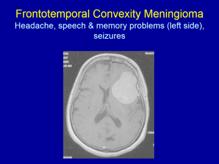
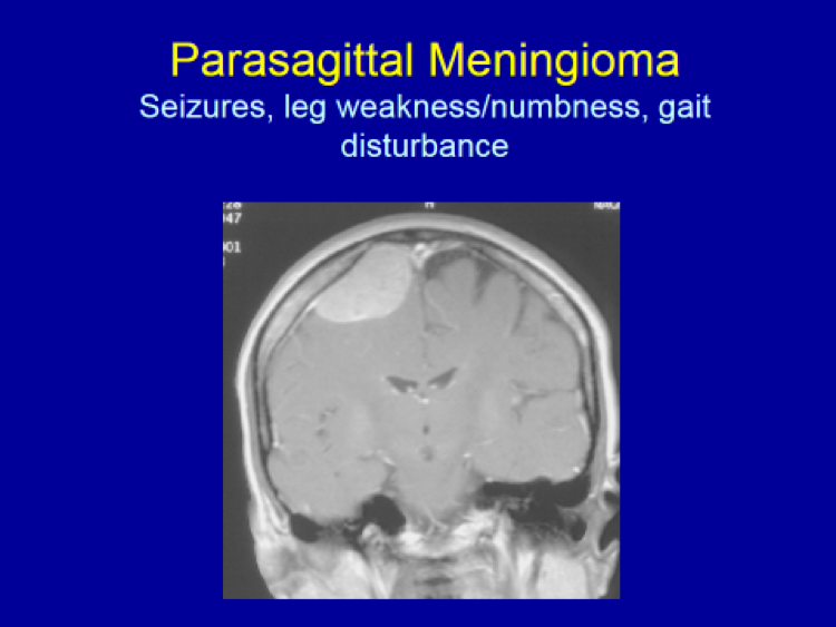
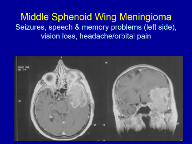
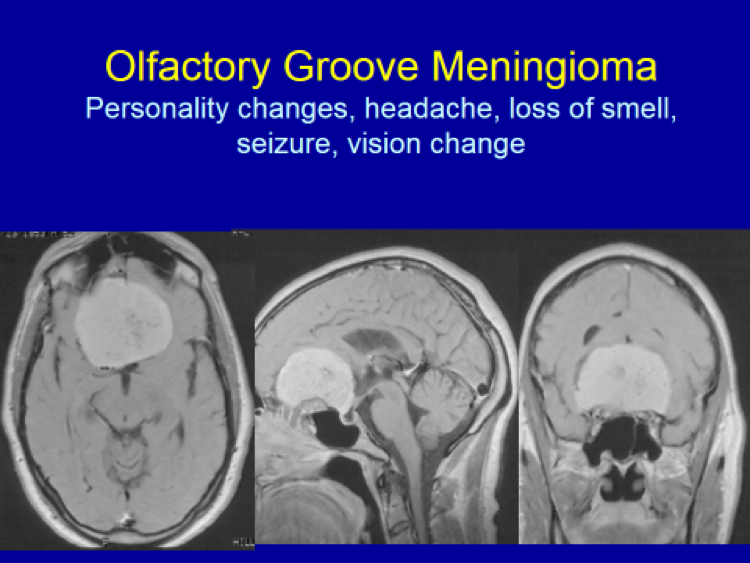
Diagnosing symptomatic meningiomas:  Brain MRI (Magnetic Resonance Imaging) is the gold standard for diagnosing intracranial meningiomas.  Usually, when patients develop any neurological symptoms – such as headaches, speech problems, seizures, memory loss, body/extremity numbness or weakness, hearing loss, visual loss, double vision, dizziness, imbalance, etc. – a brain imaging is ordered by the patient’s PCP (primary care physician).  Preferably, when a brain tumor is suspected, MRI with and without Godolinium (contrast injected intravenously) is performed.

When patients have any metal implants in the body such as cardiac pacemaker, MRI cannot be done.  Rather, a brain CT with and without IV contrast is performed.  CT is also done as an adjunct to the brain MRI when the meningioma extends into the surrounding skull or the skull base to better delineate the full extent of the tumor including the extent of bone invasion.

When meningiomas are adjacent to or involving the major venous structures, such as the sagittal sinus, torcular, transverse sinus or sigmoid sinus, MRV (Magnetic Resonance Venogram) may be performed additionally to better assess the full extent of the meningioma’s invasion into the surrounding venous structure(s).

When a very vascular meningioma (i.e. rich in blood supply) is suspected based on the preoperative MR imaging, a cerebral angiography may be performed to visualize the full extent of the blood supply to the meningioma as a part of preoperative planning.  When it would be deemed appropriate and beneficial, at the same time as the cerebral angiography is being done, tumor embolization may be performed to occlude the feeding blood vessels which, in turn, will make the subsequent surgery less bloody and risky.

Diagnosing asymptomatic, or “incidental”, meningiomas:  These days, with the widespread availability and use of brain imaging (either CT or MRI) for evaluating head trauma, nasal sinus complaints or nonspecific neurological complaints, incidental meningiomas (i.e. those detected by an “accident” or not causing any specific symptoms referable to the tumor) are being diagnosed with increasing frequency.  When a non-contrast CT done initially to evaluate nasal sinusitis or minor head trauma, for example, reveals a possible meningioma, a brain MRI with and without Gadolinium is ordered to confirm and better delineate the tumor.

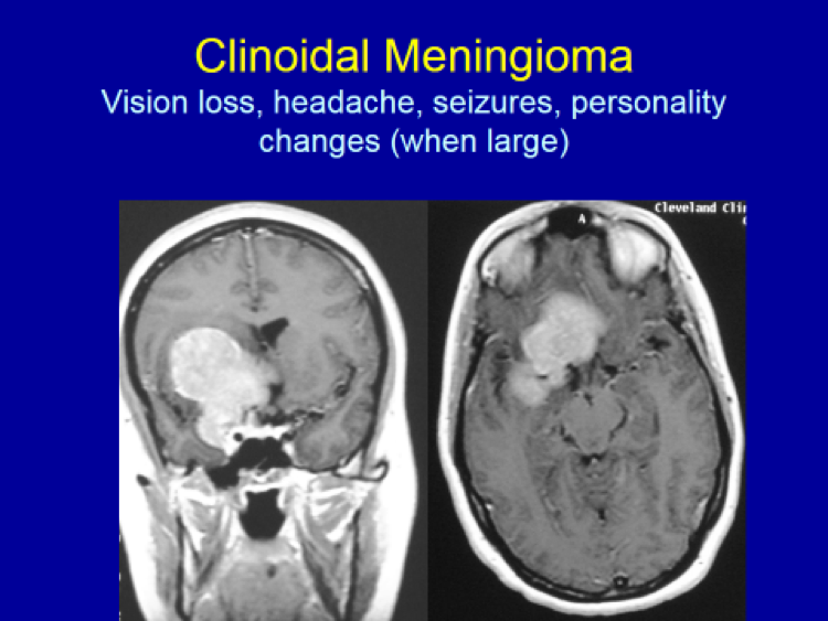
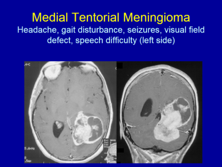
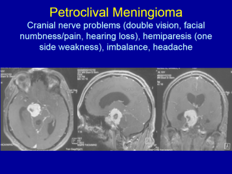
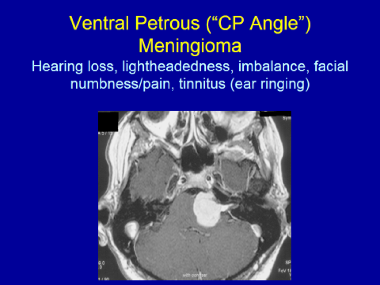
Please see the next section, “Clinical Presentation/Meningiomas by Location”, for examples of meningioma MR images.


Etiology (What’s the cause?) – So far, there are only 2 known definitive factors that lead to meningioma development:  (1) radiation, and (2) the gene associated with Neurofibromatosis type 2 (NF2).  Most likely, meningiomas and other tumors in the brain and the rest of human body develop due to alterations or damages that have occurred in the patient’s gene(s) within certain chromosomes.  Exactly what genes and what kind of alterations or damages lead to meningioma formation are still unknown other than the NF2 gene mentioned above.  However, with the NF2 gene, located on Chromosome 22, the gene defect accounts for a fraction of all (non-NF2) meningioma patients (33%).  Obviously, additional research is required to further elucidate the exact and all genetic changes that lead to meningioma formation.

Additional reading:

Lee JH, Sundaram V, Stein JD, Kinney SE, Stacey DW, Golubic M:  Reduced expression of schwannomin/merlin in human sporadic meningiomas.  Neurosurgery 40:578-587 (1997)

Evans J, Jeun SS, Cowel JK, Harwalkar JA, Golubic M, Lee JH:  Molecular alterations in the NF2 gene and its protein are rarely occurring in meningothelial meningiomas:  J Neurosurgery 94:111-117, 2001


Pathology (What cell type and how aggressive?) – Meningiomas are classified using the World Health Organization (WHO) grading scale, from Grade 1 (benign) to Grade 3 (malignant).  In a single institution review conducted by my group, 92% meningiomas belonged to WHO Grade 1 (benign), while 6% was WHO Grade 2 (aggressive or atypical) and 2% WHO Grade 3 (malignant) meningiomas.  This pathologic classification is critically important in meningiomas because one of the factors that determine how well patients do long-term depends on this tumor “grade”.  For example, only 3% of WHO Grade 1 tumors recur within 5 years of initial surgery, compared to 40% 5-year recurrence rate for WHO Grade 2 tumors, and 80% for WHO Grade 3 meningiomas.

Additional reading:

Meningothelioma as the Predominant Histological Subtype of Midline Skull Base and Spinal Meningioma

Lee JH, Sade B, Choi E, Prayson R, Golubic M:  Meningothelioma as the predominant histologic subtype of midline skull base and spinal meningiomas.  J Neurosurgery 105:60-64, 2006

Prayson R:  Pathology of meningiomas, In: Meningiomas (JH Lee, ed), Springer-Verlag, London, 2008


Definition (What is it?) – Meningiomas are predominantly benign tumors, arising from the covering (“meninges”) of the brain (90%) and spine (10%).  These tumors arise from the arachnoid “cap cells” which are the cells forming the outer lining of the arachnoid layer (the middle of the three meningeal layers).  Strictly speaking, these are not “brain tumors” per se as they do not take their origin from the brain tissue proper.

Epidemiology (How common is it?) – The incidence of symptomatic meningiomas (“how common?”)  is approximately 2/100,000 population per year while the incidence of asymptomatic (tumors causing no symptoms) meningiomas is 5.7/100,000 population per year.  Therefore, the overall combined incidence of all meningiomas (both symptomatic and asymptomatic) is nearly 8/100,000 population/year.  This roughly translates into the yearly incidence of roughly 800 new meningioma patients in a geographic area the size of Los Angeles County which has a total population of 10 million.  Symptomatic meningiomas account for about 15-20% of all primary brain tumors, second most common after glioblastoma multiforme (GBM).  When incidental (i.e. asymptomatic) tumors are included, however, meningiomas account for nearly 60% of all primary brain tumors.  In reality, meningiomas are the most common primary “brain tumors”.

The average age at diagnosis for those requiring surgery, based on the review of my personal operative series of 1200 cases, is 54 years.  For some unknown reason, meningiomas are more common in women by a ratio of 2-2.5:1.

Additional reading:

Meningioma (JH Lee, ed.), Springer-Verlag, London, 2008


© 2024 Valley Neurosurgical Institute