Meningioma Location Outcome

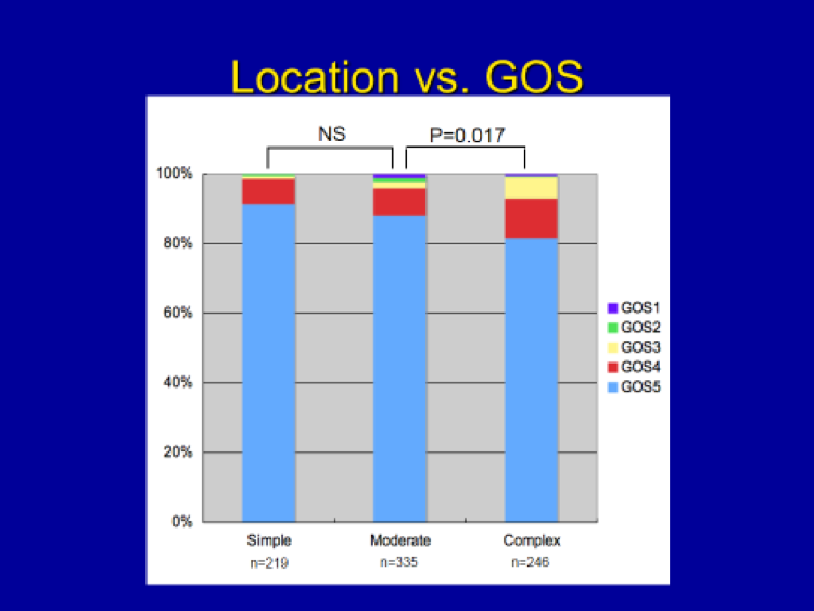
Location was analyzed with respect to outcome (GOS) by dividing the patients into 3 groups according to the level of technical difficulty posed by the tumor location (based on a review of 800 operative cases, personal series):

Simple:  Convexity, lateral sphenoid wing, cerebellar convexity, posterior petrous, posterolateral foramen magnum

Moderate:  Falx, parasagittal, middle sphenoid wing, olfactory groove, lateral tentorial,  Transverse/sigmoid sinus

Complex:  Anterior clinoid, tuberculum sella, cavernous sinus, petroclival, ventral petrous, medial tentorial, dorsal clinoid, pineal, ventral foramen magnum

No death was encountered in the “Simple” location group, and the overall incidence of excellent outcome (GOS 4 & 5) can be seen to be decreasing in a step-wise fashion going from the “Simple” to “Moderate” locations, then going from the “Moderate” locations to the “Complex” locations.

In summary, patients with meningiomas in “simple” locations do very well following surgery compared to those with tumors in more difficult or “complex” locations.  Location does matter in determining the outcome following meningioma surgery.


Co-Morbidity vs GOS

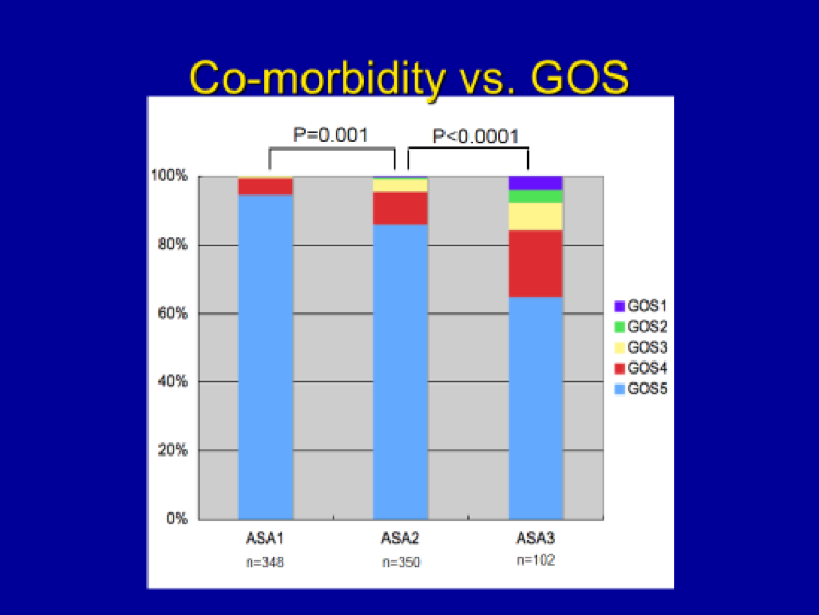
Co-morbidity (i.e. presence of any medical illnesses coexisting with meningioma at the time of surgery) was analyzed respect to outcome (GOS) using the commonly used ASA (American Society of Anesthesiologists) Physical Status Classification (ASA I – Healthy, ASA II – Mild systemic disease, ASA III – Severe systemic disease, ASA IV – Severe systemic disease that is constant threat to life, ASA IV – Moribund).   Patients with ASA Classification IV or V are not candidates for meningioma surgery as they are too ill to undergo a major operation.  (Based on a review of 800 operative cases, personal series)

Healthy patients did much better than those with significant medical illness(es).  For example, no patient in the ASA Groups I or II died after meningioma surgery.  Death was encountered only in the ASA III patients.  Additionally, the incidence of excellent outcome (GOS 4 & 5, close to 100% in the ASA Group I) decreased in a step-wise fashion in going from the ASA 1 to the ASA 2 group, and subsequently from the ASA 2 to the ASA 3 groups.

In summary, healthy patients do very well following meningioma surgery.


Based on a review of 800 personal cases of meningioma surgery as mentioned in the last section, this time we did another study in order to determine the factors that influence outcome in meningioma surgery.  In summary these important factors include the following:

Internal factor (inherent to patients/tumors):

Co-morbidity (patient’s health status)

Location

Age

Size

Symptoms

Prior surgery

Prior radiation

External factor:

Surgeon’s experience (i.e. the “learning curve” effect)

Using the first letters of the first five factors (Co-morbidity, Location, Age, Size, Symptoms), I have come up with an acronym – “CLASS”- for teaching residents and young neurosurgeons the important factors that determine outcome in meningioma surgery.  Also, utilizing these outcome factors, I have developed the “CLASS” algorithmic scale for patient selection in meningioma surgery.


Additional reading:

Factors Influencing Outcome in Meningioma Surgery

The Novel CLASS Algorithmic Scale for Patient Selection

Sade B, Lee JH:  Factors Influencing Outcome in Meningioma Surgery.  In:  “Meningiomas” (JH Lee, ed.), pp. 213-216, Springer, London, 2008

Lee JH, Sade B:  The Novel “CLASS” Algorithmic Scale for Patient Selection in Meningioma Surgery.  In:  “Meningiomas” (JH Lee, ed.), pp. 217-221, Springer, London, 2008


I have reviewed my personal operative series of 800 consecutive cases performed at the Cleveland Clinic where I had a 21-year tenure as Professor & Director, Skull Base Surgery Center, and this study represent the largest of its kind to date.  (Prior study based on 600 cases was published in my book, Meningiomas (JH Lee, ed.), see reference below.)

How patients were doing 6 weeks after their surgery was analyzed using Glasgow Outcome Scale, a common outcome measure used by neurosurgeons:

GOS I – Dead

GOS II – Vegetative

GOS III – Dependent

GOS IV – Independent

GOS V – “Normal” life, returned to preoperative employment

GOS I-III are considered as “poor” outcome, while GOS IV-V represent “good” outcome.

At 6 weeks, the following is the outcome of my 800 meningioma patients:

GOS I:    5 (0.6%)

GOS II:   6 (0.8%)

GOS III:  23 (2.9%)


Total “Poor” outcome (GOS I-III): 34 (4.3%)

GOS IV:  70 (8.8%)

GOS V:  696 (87%)


Total “Good” outcome (GOS IV-V): 766 (95.7%)


Additional reading:

Operative Outcome Following Meningioma Surgery: A Personal Experience of 600 Cases

Lee JH, Sade B:  Operative Outcome Following Meningioma Surgery:  A Personal Experience of 600 Cases.  In: “Meningiomas” (JH Lee, ed.), pp. 209-212, Springer, London, 2008


Risks involved in meningioma surgery include:

  1. Infection (1%)
  2. Excessive bleeding during surgery requiring transfusion (1-3 %, depending on the tumor size and the extent/severity of tumor blood supply)
  3. Postoperative bleeding in or around the brain surgery site, requiring re-operation (<1%)
  4. Seizures (3-5%, associated with any brain surgery other than posterior fossa surgery)
  5. Stroke (1-3%, depending on the tumor size, location, adherence to the surrounding blood vessel)
    1. Symptoms/deficits depend on the extent and location of stroke
  6. Direct brain injury or worsening postoperative brain swelling (extremely rare, <<1%)
    1. Symptoms/deficits depend on the extent and location of brain injury
  7. Cranial nerve injury (1-3%, depending on the tumor size, location, adherence to the surrounding cranial nerves)
    1. Symptoms/deficits depend on the specific cranial nerves involved:
    2. Loss of smell, blindness, double vision, facial numbness, hearing loss, facial weakness, hoarseness, swallowing difficulty, weak cough, tongue/shoulder weakness
    3. CSF (cerebrospinal fluid) leak (depending on the tumor’s skull base location, <1%-3%)
  8. Inflammatory reaction (“chemical meningitis”) to an artificial dural graft when used (2-3% of patients with artificial dural graft usage)
  9. Persistent postoperative headache around the incision (rare, ~1%)
  10. Postoperative medical problems (1-3%, depending on the patient’s age, health, length of surgery, postoperative mobility)
    1. Urinary tract infection
    2. Pneumonia
    3. Deep venous thrombosis (DVT)/pulmonary embolism (PE)
    4. Myocardial infarction (MI)


Meningiomas of different locations require varying surgical approaches that are primarily dictated by anatomic considerations inherent to each particular tumor location.  Additional factors that lead to variations in surgical approach include the surgeon’s personal experience and preference.

The following basic principles hold for meningioma surgery of most locations:

  1. Optimal patient positioning and incision
  1. Craniotomy (skull opening), dural opening and the tumor exposure
  1. Early tumor devascularizaion (“cutting off the blood supply to the tumor”)
  1. Internal decompression and extracapsular dissection
  1. Early localization and preservation of adherent or adjacent neurovascular structures (the brain tissue, cranial nerves, blood vessels)
  1. Removal of the involved bone and dura
  1. Optimal dural reconstruction and operative wound closure


Additional reading:

Management Options and Surgical Principles: An Overview

Surgical Management of Convexity Meningiomas

Surgical Technique for Removal of Clinoidal Meningiomas

Lee JH, Sade B:  Management Options and Surgical Principles:  An Overview.  In:  “Meningiomas” (JH Lee, ed.), pp. 203-207, Springer, London, 2008

Steinmetz MP, Krishnaney A, Lee JH:  Surgical Management of Convexity Meningiomas.  In:  Neurosurgical Operative Atlas – Neuro-Oncology (Badie B, ed.), pp. 145-152, Thieme, NYC, 2007

Lee JH, Evans JJ, Steinmetz MP, Kwon JT.  Surgical Technique for Removal of Clinoidal Meningiomas.  In:  Neurosurgical Operative Atlas – Neuro-Oncology (Badie B., ed.), pp. 153-160, Thieme, NYC, 2007

Oya S, Sade B, Lee JH:  Spheno-orbital meningioma:  Surgical Technique and Outcome.  J Neurosurg 114:1241-1249, 2011


  1. Patients with neurological symptoms and/or deficits
  1. Patients with large (>3-4 cm) tumors
  1. Young patients (<60) with any size tumors
  1. Patients with tumors with no calcification, T2 hyperintensity, significant peritumoral edema (see the “Natural History” Section)
  1. Patients with tumors in locations carrying reasonable surgical risks
  1. Patients who are healthy
  1. Patients with tumors which grew after a period of initial observation
  1. Patients favoring surgery over observation or radiation after thorough discussion


I write (note the present tense as this is a work in progress which will be updated from time to time) this short “Handbook” mainly as a small part in my “giving back” for abundant blessings I have received throughout my life.

First and foremost, I thank God for my life, faith, family, and career that I love.  I feel so blessed for living my childhood dream, which was to become a neurosurgeon.  (Please see “Living my childhood dream” under my personal blog section.)  When I go to work each day, I do so with tremendous excitement and gratitude, because I truly love what I do.  Looking back at my life, God has always guided me along the best possible path and blessed me richly that led to the present.

Second, I have always felt a great sense of gratitude towards all my former patients who taught me so much about the profession I love.  Two of the most important personal principles of mine in medicine are (1) to treat each patient as family, and (2) to regard each patient as a teacher.  It is this second principle that motivated me throughout my career to conduct research studies, write academic papers, and learn from each of my patients so that my future patients will receive better and improved care.  This handbook is a small token of my deep appreciation to all my patients – my real teachers.

I also write this handbook as a token of sincere apology to my former patients.  I am truly sorry that I did not have this available to them at the time of their seeing me for their meningiomas.  I felt the need for this type of simple handbook a long time ago, but I just did not find the time to do it sooner.  Many of my former patients and their family members often spent hours, with significant anxiety and fear of having been diagnosed with a “meningioma”, doing “Google” search for a word that was difficult even to pronounce.  More often than not, having difficulty finding anything adequate and simple, they spent hours perusing scientific articles (with a dictionary by their side) written in a language so foreign to them.

It is my wish and intention that this handbook will give meningioma patients all the necessary basic information about their newly diagnosed condition.  I want to empower them with knowledge about their “brain tumor” so that their anxiety is eased, and they are better equipped to make educated treatment decisions.

It is also my great wish that the facts outlined in this handbook will give them hope.  Rather than asking “Why me?” after receiving the diagnosis, I want all those who read this to finish the handbook on a positive note, having hope that meningiomas can be “cured” or, at least, put under “control” without significantly altering their life quality or life expectancy.

Before closing, I thank my mentor, the late Dr. John A. Jane, Sr., for teaching me and equipping me so that I can live my dream as a neurosurgeon.  His spirit lives on in my drive, passion and love for neurosurgery.  Also, I thank my lovely wife of 31 years for her constant love, support and inspiration.  She has been by my side from the very beginning of my journey as a neurosurgeon, for we got married one month before the start of my residency.  Lastly, I thank my 93-year old mother whose daily prayers and love shaped me as a person.  She was the main reason for me to return “home” to L.A. in 2014 so that I can spend some quality time with her in the remaining few years of her life.


I just do not feel “right” to be charging any money for this Handbook as it represents a final product from labor of love – for neurosurgery, my patients and the topic of meningioma – motivated by a strong sense of indebtedness and appreciation to all of my former patients for everything they taught me.

As you read this Handbook, please take a moment to thank those who came before you, without whom this Handbook and the improved care I am now able to provide today would not be possible.

Rather than “purchasing”, which would greatly cheapen the true value of their individual contributions to my neurosurgical “education and training”, you may download this entire handbook (or any part of it) in any of the following 4 ways:

  • Making a “Special Offering of Thanks” to your church in any amount,
  • Making a donation to any charitable organization of your choice,
  • Sharing the link for this Handbook (ValleyNI.com) with any friends, relatives or acquaintances with meningioma who can benefit from it,
  • Making a donation, in any amount, to our non-profit foundation, “Equal Care Foundation”, which helps to provide the same world-class neurosurgical care to all patients regardless of their financial ability or background(Pay to the order of “Equal Care Foundation”).
    Equal Care Foundation
    c/o Valley Neurosurgical Institute
    501 South Buena Vista Street,
    Burbank, CA  91505

With best wishes for your winning the battle with meningioma,

Joung H. Lee, M.D.
President, Valley Neurosurgical Institute &
Co-Director, Hollywood Presbyterian Neuroscience Institute

www.ValleyNI.com


Surgery is the treatment of choice for most patients with meningiomas.  In patients with benign meningiomas (WHO Grade I, comprising 92% of all meningiomas), the tumor location largely dictates the extent of removal, which, in turn determines the tumor recurrence and, ultimately, the patient’s long-term progression-free survival.

Primary goals of surgery include, as alluded to in the prior section (“Surgery:  Indication and Goals”):  (1) total removal of the tumor and the involved surrounding bone and dura (the brain covering from which meningiomas arise) when feasible, thereby possibly providing cure or significantly altering the natural history of the disease process, and (2) reversal or improvement in neurologic deficits/symptoms caused by the tumor.

In meningiomas of certain locations that are associated with very high operative morbidity, such as the cavernous sinus or petroclival regions where complete resection is not always possible, additional surgical goals may include confirmation of the tissue diagnosis and tumor reduction to less than 3 cm in maximum diameter in preparation for postoperative radiosurgery.

Given the benign nature of meningiomas and the established efficacy of adjuvant radiation, the goal of total removal must be balanced by the physician’s basic credo to “do no harm”.  When total removal carries a significant risk of morbidity, a small piece of tumor may be left, with further plans of observation followed by reoperation or radiation when the tumor is noted to be growing or causing new symptoms.

Operative Goals (Summary):

  1. When possible, total removal including the surrounding/involved dura (the brain covering from which meningiomas arise) and the bone (skull), to provide “surgical cure”. This type of extensive surgery insures significantly altered natural history of the meningioma, and thereby prolonging one’s life (Benefit 1 as discussed in the “Management:  Basic Principles” Section, #4.)
  1. Improvement and preservation of neurological function, thereby providing patients with optimal life quality. (Benefit 2)
  1. Planned subtotal removal for patients in whom total resection carries significant risks of morbidity.
  1. In short, it is critically important to remove “as much as possible and, at the same time, as safely as possible.” Knowing when to stop during meningioma surgery comes from the surgeon’s wisdom, gained through years of experience.

© 2024 Valley Neurosurgical Institute