Risks involved in meningioma surgery include:

  1. Infection (1%)
  2. Excessive bleeding during surgery requiring transfusion (1-3 %, depending on the tumor size and the extent/severity of tumor blood supply)
  3. Postoperative bleeding in or around the brain surgery site, requiring re-operation (<1%)
  4. Seizures (3-5%, associated with any brain surgery other than posterior fossa surgery)
  5. Stroke (1-3%, depending on the tumor size, location, adherence to the surrounding blood vessel)
    1. Symptoms/deficits depend on the extent and location of stroke
  6. Direct brain injury or worsening postoperative brain swelling (extremely rare, <<1%)
    1. Symptoms/deficits depend on the extent and location of brain injury
  7. Cranial nerve injury (1-3%, depending on the tumor size, location, adherence to the surrounding cranial nerves)
    1. Symptoms/deficits depend on the specific cranial nerves involved:
    2. Loss of smell, blindness, double vision, facial numbness, hearing loss, facial weakness, hoarseness, swallowing difficulty, weak cough, tongue/shoulder weakness
    3. CSF (cerebrospinal fluid) leak (depending on the tumor’s skull base location, <1%-3%)
  8. Inflammatory reaction (“chemical meningitis”) to an artificial dural graft when used (2-3% of patients with artificial dural graft usage)
  9. Persistent postoperative headache around the incision (rare, ~1%)
  10. Postoperative medical problems (1-3%, depending on the patient’s age, health, length of surgery, postoperative mobility)
    1. Urinary tract infection
    2. Pneumonia
    3. Deep venous thrombosis (DVT)/pulmonary embolism (PE)
    4. Myocardial infarction (MI)


Meningiomas of different locations require varying surgical approaches that are primarily dictated by anatomic considerations inherent to each particular tumor location.  Additional factors that lead to variations in surgical approach include the surgeon’s personal experience and preference.

The following basic principles hold for meningioma surgery of most locations:

  1. Optimal patient positioning and incision
  1. Craniotomy (skull opening), dural opening and the tumor exposure
  1. Early tumor devascularizaion (“cutting off the blood supply to the tumor”)
  1. Internal decompression and extracapsular dissection
  1. Early localization and preservation of adherent or adjacent neurovascular structures (the brain tissue, cranial nerves, blood vessels)
  1. Removal of the involved bone and dura
  1. Optimal dural reconstruction and operative wound closure


Additional reading:

Management Options and Surgical Principles: An Overview

Surgical Management of Convexity Meningiomas

Surgical Technique for Removal of Clinoidal Meningiomas

Lee JH, Sade B:  Management Options and Surgical Principles:  An Overview.  In:  “Meningiomas” (JH Lee, ed.), pp. 203-207, Springer, London, 2008

Steinmetz MP, Krishnaney A, Lee JH:  Surgical Management of Convexity Meningiomas.  In:  Neurosurgical Operative Atlas – Neuro-Oncology (Badie B, ed.), pp. 145-152, Thieme, NYC, 2007

Lee JH, Evans JJ, Steinmetz MP, Kwon JT.  Surgical Technique for Removal of Clinoidal Meningiomas.  In:  Neurosurgical Operative Atlas – Neuro-Oncology (Badie B., ed.), pp. 153-160, Thieme, NYC, 2007

Oya S, Sade B, Lee JH:  Spheno-orbital meningioma:  Surgical Technique and Outcome.  J Neurosurg 114:1241-1249, 2011


  1. Patients with neurological symptoms and/or deficits
  1. Patients with large (>3-4 cm) tumors
  1. Young patients (<60) with any size tumors
  1. Patients with tumors with no calcification, T2 hyperintensity, significant peritumoral edema (see the “Natural History” Section)
  1. Patients with tumors in locations carrying reasonable surgical risks
  1. Patients who are healthy
  1. Patients with tumors which grew after a period of initial observation
  1. Patients favoring surgery over observation or radiation after thorough discussion


I write (note the present tense as this is a work in progress which will be updated from time to time) this short “Handbook” mainly as a small part in my “giving back” for abundant blessings I have received throughout my life.

First and foremost, I thank God for my life, faith, family, and career that I love.  I feel so blessed for living my childhood dream, which was to become a neurosurgeon.  (Please see “Living my childhood dream” under my personal blog section.)  When I go to work each day, I do so with tremendous excitement and gratitude, because I truly love what I do.  Looking back at my life, God has always guided me along the best possible path and blessed me richly that led to the present.

Second, I have always felt a great sense of gratitude towards all my former patients who taught me so much about the profession I love.  Two of the most important personal principles of mine in medicine are (1) to treat each patient as family, and (2) to regard each patient as a teacher.  It is this second principle that motivated me throughout my career to conduct research studies, write academic papers, and learn from each of my patients so that my future patients will receive better and improved care.  This handbook is a small token of my deep appreciation to all my patients – my real teachers.

I also write this handbook as a token of sincere apology to my former patients.  I am truly sorry that I did not have this available to them at the time of their seeing me for their meningiomas.  I felt the need for this type of simple handbook a long time ago, but I just did not find the time to do it sooner.  Many of my former patients and their family members often spent hours, with significant anxiety and fear of having been diagnosed with a “meningioma”, doing “Google” search for a word that was difficult even to pronounce.  More often than not, having difficulty finding anything adequate and simple, they spent hours perusing scientific articles (with a dictionary by their side) written in a language so foreign to them.

It is my wish and intention that this handbook will give meningioma patients all the necessary basic information about their newly diagnosed condition.  I want to empower them with knowledge about their “brain tumor” so that their anxiety is eased, and they are better equipped to make educated treatment decisions.

It is also my great wish that the facts outlined in this handbook will give them hope.  Rather than asking “Why me?” after receiving the diagnosis, I want all those who read this to finish the handbook on a positive note, having hope that meningiomas can be “cured” or, at least, put under “control” without significantly altering their life quality or life expectancy.

Before closing, I thank my mentor, the late Dr. John A. Jane, Sr., for teaching me and equipping me so that I can live my dream as a neurosurgeon.  His spirit lives on in my drive, passion and love for neurosurgery.  Also, I thank my lovely wife of 31 years for her constant love, support and inspiration.  She has been by my side from the very beginning of my journey as a neurosurgeon, for we got married one month before the start of my residency.  Lastly, I thank my 93-year old mother whose daily prayers and love shaped me as a person.  She was the main reason for me to return “home” to L.A. in 2014 so that I can spend some quality time with her in the remaining few years of her life.


I just do not feel “right” to be charging any money for this Handbook as it represents a final product from labor of love – for neurosurgery, my patients and the topic of meningioma – motivated by a strong sense of indebtedness and appreciation to all of my former patients for everything they taught me.

As you read this Handbook, please take a moment to thank those who came before you, without whom this Handbook and the improved care I am now able to provide today would not be possible.

Rather than “purchasing”, which would greatly cheapen the true value of their individual contributions to my neurosurgical “education and training”, you may download this entire handbook (or any part of it) in any of the following 4 ways:

  • Making a “Special Offering of Thanks” to your church in any amount,
  • Making a donation to any charitable organization of your choice,
  • Sharing the link for this Handbook (ValleyNI.com) with any friends, relatives or acquaintances with meningioma who can benefit from it,
  • Making a donation, in any amount, to our non-profit foundation, “Equal Care Foundation”, which helps to provide the same world-class neurosurgical care to all patients regardless of their financial ability or background(Pay to the order of “Equal Care Foundation”).
    Equal Care Foundation
    c/o Valley Neurosurgical Institute
    501 South Buena Vista Street,
    Burbank, CA  91505

With best wishes for your winning the battle with meningioma,

Joung H. Lee, M.D.
President, Valley Neurosurgical Institute &
Co-Director, Hollywood Presbyterian Neuroscience Institute

www.ValleyNI.com


Surgery is the treatment of choice for most patients with meningiomas.  In patients with benign meningiomas (WHO Grade I, comprising 92% of all meningiomas), the tumor location largely dictates the extent of removal, which, in turn determines the tumor recurrence and, ultimately, the patient’s long-term progression-free survival.

Primary goals of surgery include, as alluded to in the prior section (“Surgery:  Indication and Goals”):  (1) total removal of the tumor and the involved surrounding bone and dura (the brain covering from which meningiomas arise) when feasible, thereby possibly providing cure or significantly altering the natural history of the disease process, and (2) reversal or improvement in neurologic deficits/symptoms caused by the tumor.

In meningiomas of certain locations that are associated with very high operative morbidity, such as the cavernous sinus or petroclival regions where complete resection is not always possible, additional surgical goals may include confirmation of the tissue diagnosis and tumor reduction to less than 3 cm in maximum diameter in preparation for postoperative radiosurgery.

Given the benign nature of meningiomas and the established efficacy of adjuvant radiation, the goal of total removal must be balanced by the physician’s basic credo to “do no harm”.  When total removal carries a significant risk of morbidity, a small piece of tumor may be left, with further plans of observation followed by reoperation or radiation when the tumor is noted to be growing or causing new symptoms.

Operative Goals (Summary):

  1. When possible, total removal including the surrounding/involved dura (the brain covering from which meningiomas arise) and the bone (skull), to provide “surgical cure”. This type of extensive surgery insures significantly altered natural history of the meningioma, and thereby prolonging one’s life (Benefit 1 as discussed in the “Management:  Basic Principles” Section, #4.)
  1. Improvement and preservation of neurological function, thereby providing patients with optimal life quality. (Benefit 2)
  1. Planned subtotal removal for patients in whom total resection carries significant risks of morbidity.
  1. In short, it is critically important to remove “as much as possible and, at the same time, as safely as possible.” Knowing when to stop during meningioma surgery comes from the surgeon’s wisdom, gained through years of experience.


Observation:  Indications (When to observe?)

  1. Patients with no symptoms (“incidental meningiomas”) of all ages
  1. Patients with mild symptoms in the elderly or those patients with serious medical conditions in whom surgery carries a significant risks.
  1. Patients with a long history of mild or non-progressive symptoms with tumors in the “high risk” location (e.g. hearing loss on one side)
  1. Patients who strongly oppose surgery after thorough counselling

Radiation:  Indications (When to radiate?)

  1. Patients who have residual tumors with documented growth, or with malignant tumor histology (i.e. WHO Grade III)
  1. Patients with nonoperated tumors or recurrence not amenable to safe surgery (due to the patient’s medical condition, tumor location, or age)
  1. Patients who strongly favor radiation over surgery after thorough discussion of all treatment options
  1. Conventional radiation (or IMRT – ‘intensity modulated radiation therapy”) for patients with tumor size greater than 3-4 cm
  1. Radiosurgery (“Gamma Knife Radiosurgery” or “Cyber Knife Radiosurgery”) for patients with tumor size less than 3 cm


  1. As indicated in the preceding “Natural History” Section, not all meningiomas require any treatment, either in the form of surgery or radiation, at the time of initial diagnosis/presentation.  In my personal practice while at the Cleveland Clinic, among over 200 new patients with meningiomas evaluated each year, about 100 patients were treated with surgery, 10 patients with radiation (or radiosurgery) and 100 patients were observed.
  2. Treatment offered (either surgery or radiation) must guarantee an outcome that is far superior over the natural history (i.e. untreated) itself.
  3. Treatment benefits must far outweigh the risks.
  4. There are only two treatment benefits in meningioma management:
    1. Significant alteration of the natural history of the disease (“prolonging life”)
    2. Reversal or improvement of the presenting neurological symptoms/deficits (“improving life quality”)
  5. There are 3 categories of management options:
    1. Observation
    2. Surgery
    3. Radiation/Radiosurgery

Additional reading:

Lee JH & Sade B:  Management Options and Surgical Principles – An Overview (JH Lee, ed), pp. 203-207), Springer, London, 2008


The natural history of a disease (e.g. a meningioma) is the course an illness takes without any intervention (i.e. any treatment).  In other words, the natural history of a meningioma is how that particular tumor behaves in an individual patient when it is just observed without any treatment.

In any given disease, when and what treatment should be administered must be based on thorough knowledge of (1) the disease’s natural history, (2) the risks of the treatment, and (3) the benefits of the treatment.  In surgical management of a meningioma patient, benefits of surgery must far outweigh the risks, and simultaneously, surgery must significantly alter the natural history, either by “curing” the meningioma when “complete” removal is achieved, or by significantly “delaying the (tumor) progression” when “partial” removal is performed, thereby prolonging the patient’s overall survival.

The literature information on meningioma’s “natural history” has been scant, and when available, the past studies were limited to the sample size of less than 100 patients.  My group at the Cleveland Clinic published the largest “natural history” paper on meningiomas in 2011.  In that study, we observed 273 meningiomas in 244 patients for a mean period of nearly 4 years.   In that time frame, 44% of the meningiomas showed growth.  Among the factors that “favored” growth in these observed meningiomas were:  (1) young age less than 60 years old, (2) absence of calcification in meningiomas, (3) T2 signal hyperintensity on MR imaging, and (4) peritumoral edema (“brain swelling” around the tumor).

What the above study indicates are the following:

  • Not all meningiomas, especially if they are not causing any symptoms, require surgery at the time of initial diagnosis as only less than half (44%) show growth in 4 years.
  • In patients with meningiomas that have any of the above 4 factors that favor growth, surgery should be considered either at the time of diagnosis, or when the tumor shows growth after an initial period of observation.
  • If 44% of observed meningiomas showed growth in 4 years, it is reasonable to deduce that a vast majority of meningiomas will eventually grow when observed beyond 5-10 years. Therefore, in young patients, surgery may be considered at the time of diagnosis while the tumor is at its smallest size.  In brain surgery, the larger the tumor size, the higher are the surgical risks.


Additional reading:

The Natural History of Intracranial Meningiomas

Oya S, Kim SH, Sade B, Lee JH:  The natural history of intracranial meningiomas, J Neurosurg 114:1250-1256 (2011)


Meningioma may cause varying symptoms, mainly dictated by (1) the tumor location and (2) the extent of the “mass effect” or pressure exerted on the surrounding anatomic structures by the tumor itself and/or the “edema” (i.e. brain swelling) caused by the tumor when present.  Small tumors, in the absence of any severe surrounding edema, usually do not cause any significant symptoms because of the negligible “mass effect”.  Sometimes, even large tumors may not cause any significant symptoms mainly because the tumor grew slowly and the surrounding brain accommodated well the gradually progressing mass effect.

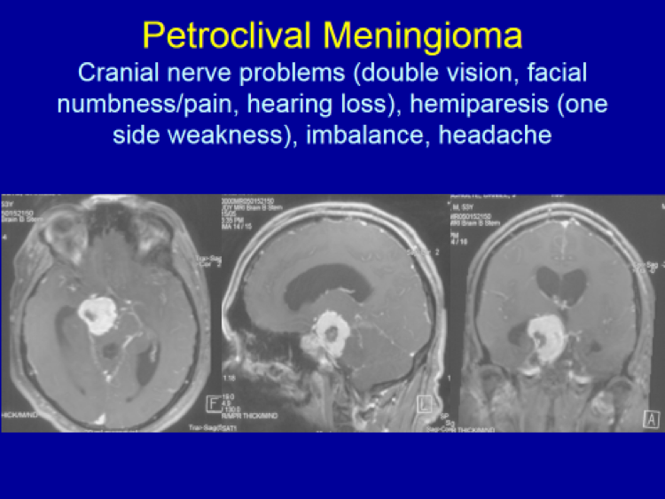
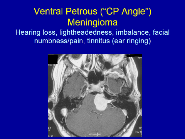
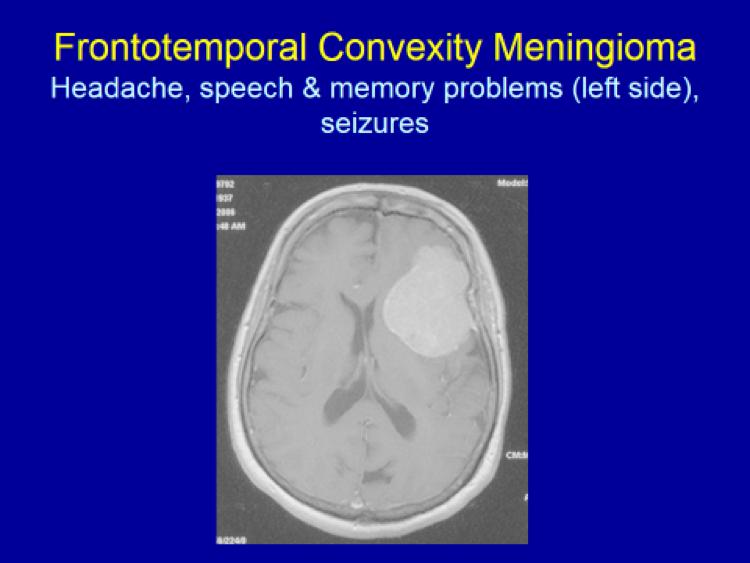
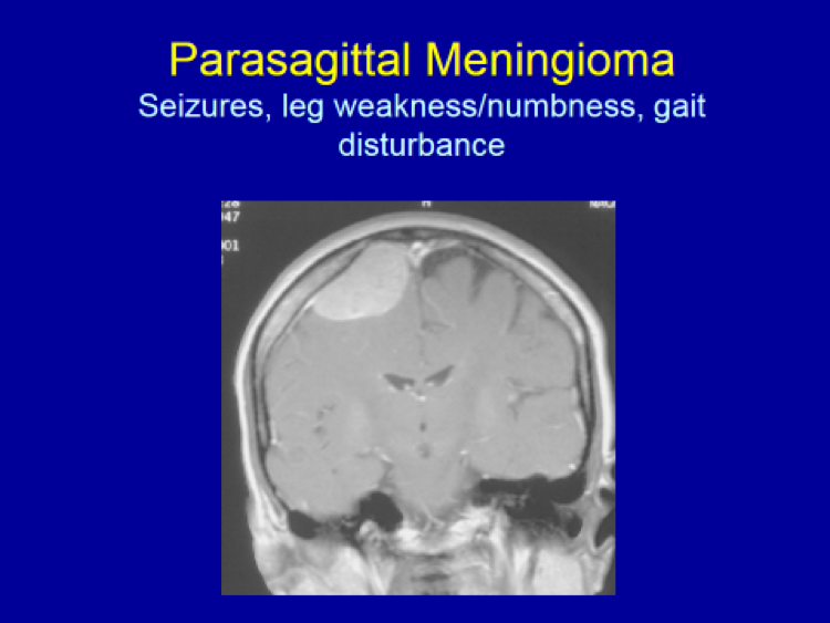
Symptoms (“clinical presentation”) caused by meningiomas are direct reflection on the particular function of the brain/cranial nerve(s) affected by the tumor’s mass effect:

Any forebrain areas (i.e. the frontal, temporal, parietal and occipital lobes) – Seizures   and headache when large

Frontal lobe – Personality changes

Left temporal lobe – Memory and/or speech problems

Frontoparietal lobe – Focal seziures, focal weakness/numbness on the opposite side

Optic nerve (CN II) – Visual deterioration

Occipital lobe – Visual field deficit

Cerebellum (the “hind brain”) – Balance and coordination problems, walking difficulty

Other cranial nerves – Loss of smell (CN I), facial pain/numbness (CN V), double vision (CN III, IV, VI), hearing loss (CN VIII), swallowing difficulty and/or hoarseness (CN X)

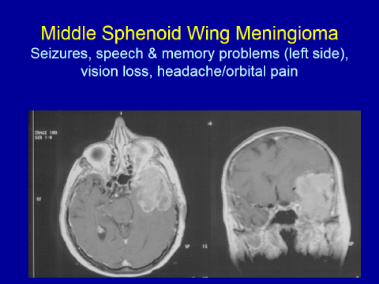
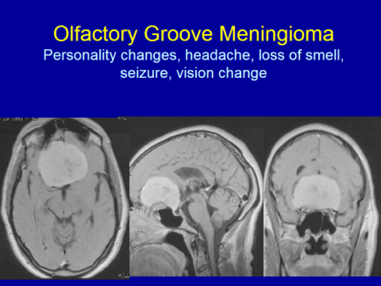
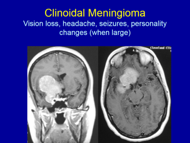
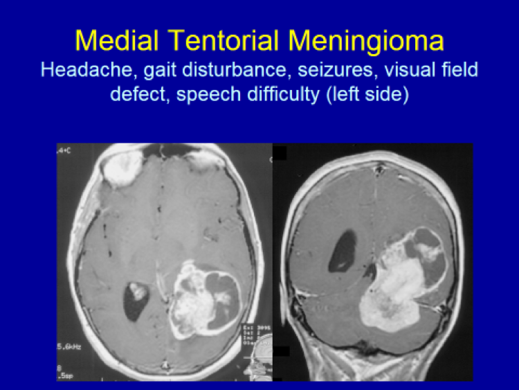
The following images are selected, and not comprehensive, examples of meningiomas.  Common symptoms caused by these meningiomas according to their locations are presented.

Clinical Presentation & Radiographic Appearance

Frontotemporal Convexity Meningioma

Parasagittal Meningioma

Middle Sphenoid Wing Meningioma

Olfactory Groove Meningioma

Clinoidal Meningioma

Medial Tentorial Meningioma

Petroclival Meningioma

Ventral Petrous Meningioma


© 2024 Valley Neurosurgical Institute